Sarpeda unlocks twice as many markers and pristine signal from your existing microscopes — no hardware changes, no new reagents. A pure software upgrade for the infrastructure you already own.
See It in ActionJust 3–4 color channels to resolve dozens of cell types, co-expression patterns, and spatial relationships. Biology is far more complex than what conventional microscopy can capture in a single acquisition.
Autofluorescence, channel bleed-through, and non-specific binding bury the true signal. Researchers constantly fight a thick wall of noise that obscures what matters most.
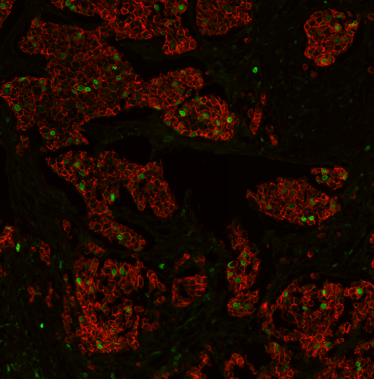
Separates two markers labeled with the same fluorophore using AI. Double your marker capacity in a single acquisition. Six markers plus DAPI on a four-channel microscope — from one tissue section with standard reagents.
Precision removal of autofluorescence, bleed-through, and non-specific binding artifacts. A true, complete separation of all background signal — learned from routine negative controls. This is not your typical background subtraction.
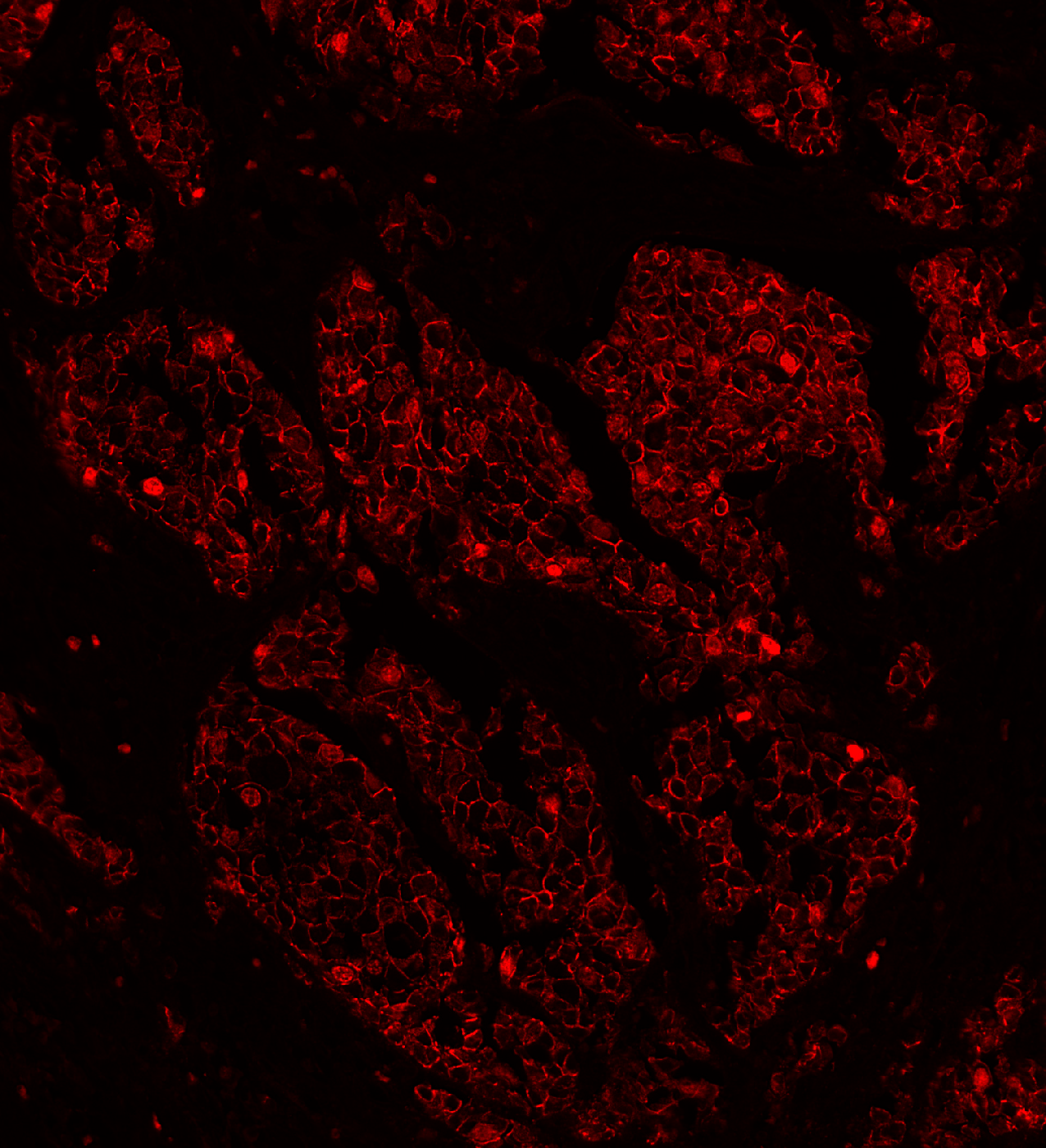
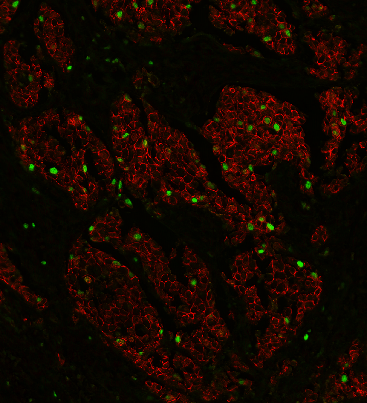
Breast tissue: E-cadherin and Ki-67 labeled with the same fluorophore, then separated by AI. Green nuclei (Ki-67) clearly emerge from the red membrane staining (E-cadherin).
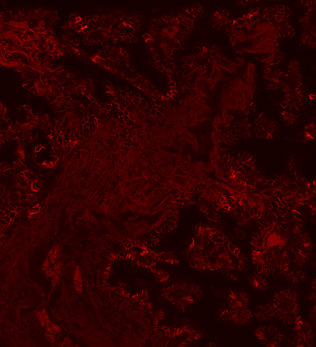
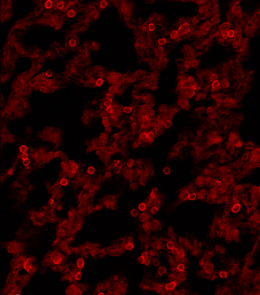
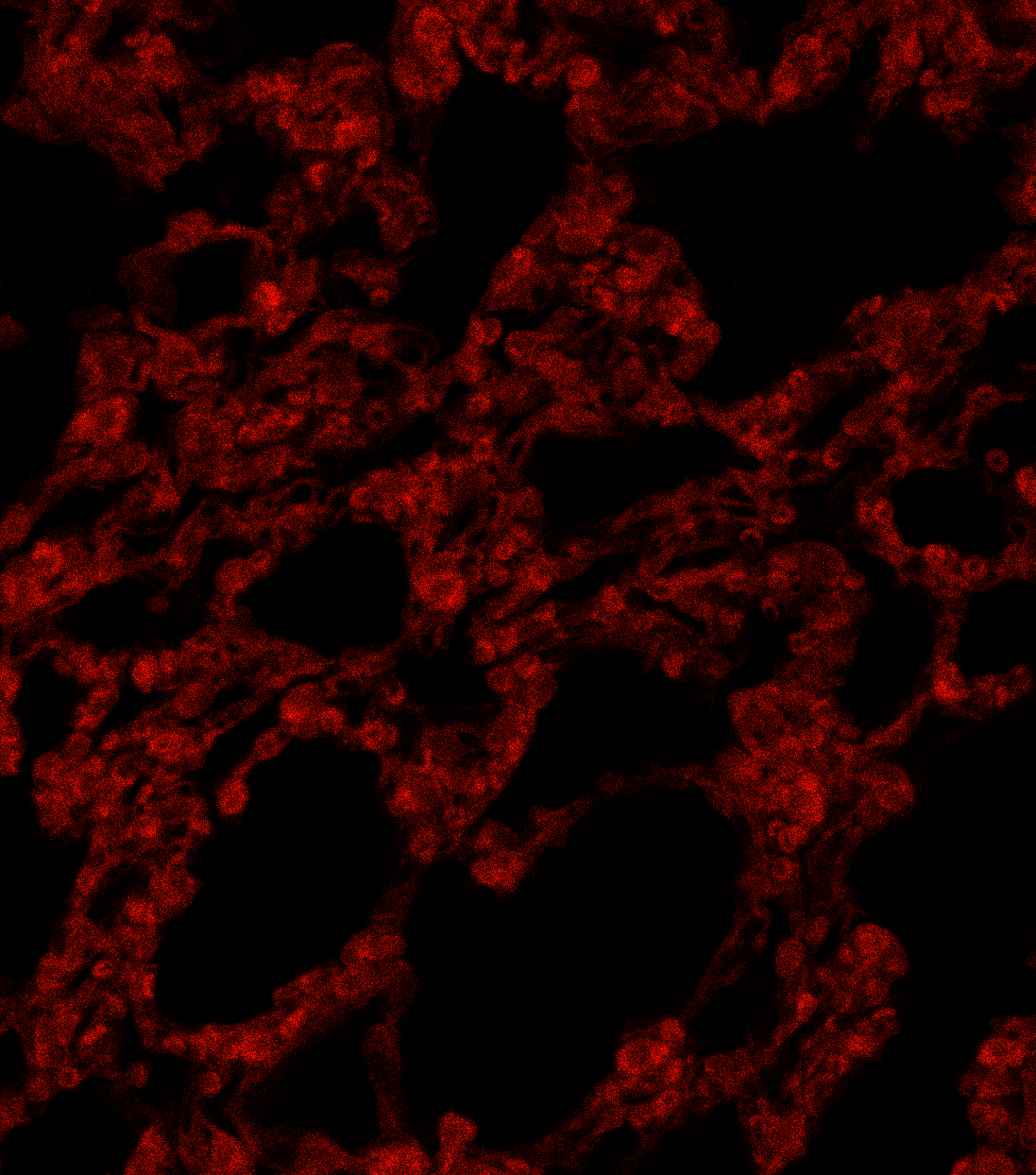
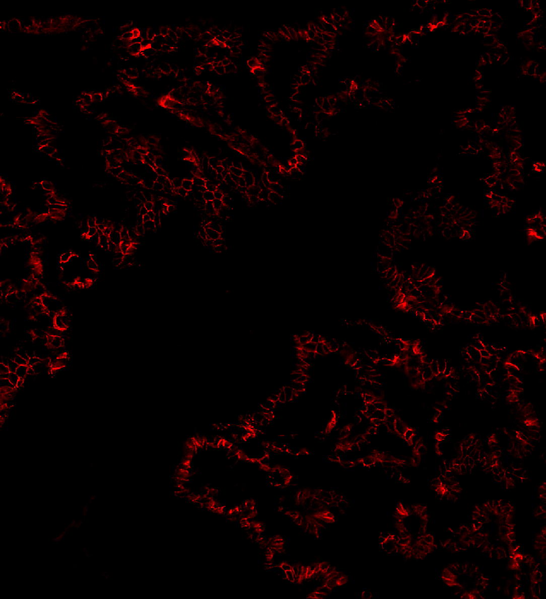
Lung tissue: E-cadherin signal buried in autofluorescence, then cleaned by AI. The true biological signal emerges with single-pixel detail preserved.
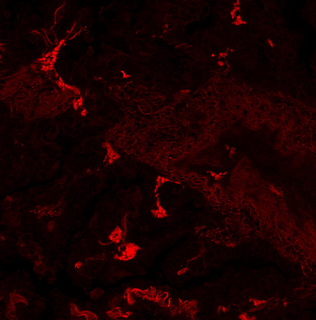
Lung tissue, 568 nm channel: the target aSMA signal is contaminated by crosstalk bleeding in from the Rb in the adjacent 647 nm channel. Sarpeda Clear removes the bleed-through and recovers the true aSMA signal.
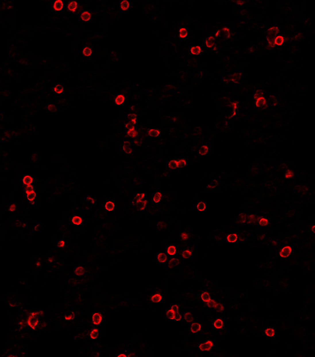
Six markers plus DAPI on a four-channel microscope, from a single tissue section. With indirect labeling, only three standard species needed — simpler reagents, lower cost.
Precision removal of autofluorescence, channel bleed-through, and non-specific binding. Reveal the true biological signal hidden beneath noise.
Works with any microscope platform. Integrates into existing analysis pipelines. No hardware changes, no new reagents. A software upgrade for existing infrastructure.
Conserve precious patient samples. Achieve true co-expression analysis in a single acquisition with doubled marker capacity.
More accurate RNA reporter detection with clean signal. Cut the number of imaging cycles in half.
Marker-rich phenotypic assays with reliable downstream quantification. Rescue failed acquisitions that would otherwise be lost.
Background removed from 3D matrices. Higher information density per sample. Fewer replicates needed.
Our CEO Ivana Mikić is giving a talk and is presenting a poster showcasing our technology. She is also participating in a panel on organoid screening scalability. The video below is looping in the exhibition hall.
Sarpeda is exhibiting in the Breakthrough Zone at ELRIG Drug Discovery 2026. Our AI team is at the stand with live demonstrations of marker separation and background removal.
Attendance is free. To reserve time with us at the show, get in touch.
Book a meeting at ELRIG →
20+ years in AI and biological imaging. Scientist, inventor and repeat founder.
20+ years in cutting-edge AI, spanning academia and industry.
Professor & Microscopy Core Director at Baylor College of Medicine. Early Sarpeda adopter.
Precision oncology and spatial biology CEO.
Principal of Luna Lifesciences. Founding CEO of AxoSim (now 28bio). Organ-on-a-chip pioneer.
We're signing up partners ready to transform what their microscopes can do.
Contact Us