See the Difference
AI-Powered Results
Drag the slider to compare input images with Sarpeda's output. The difference speaks for itself.
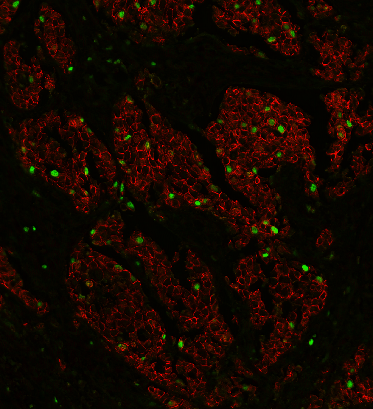
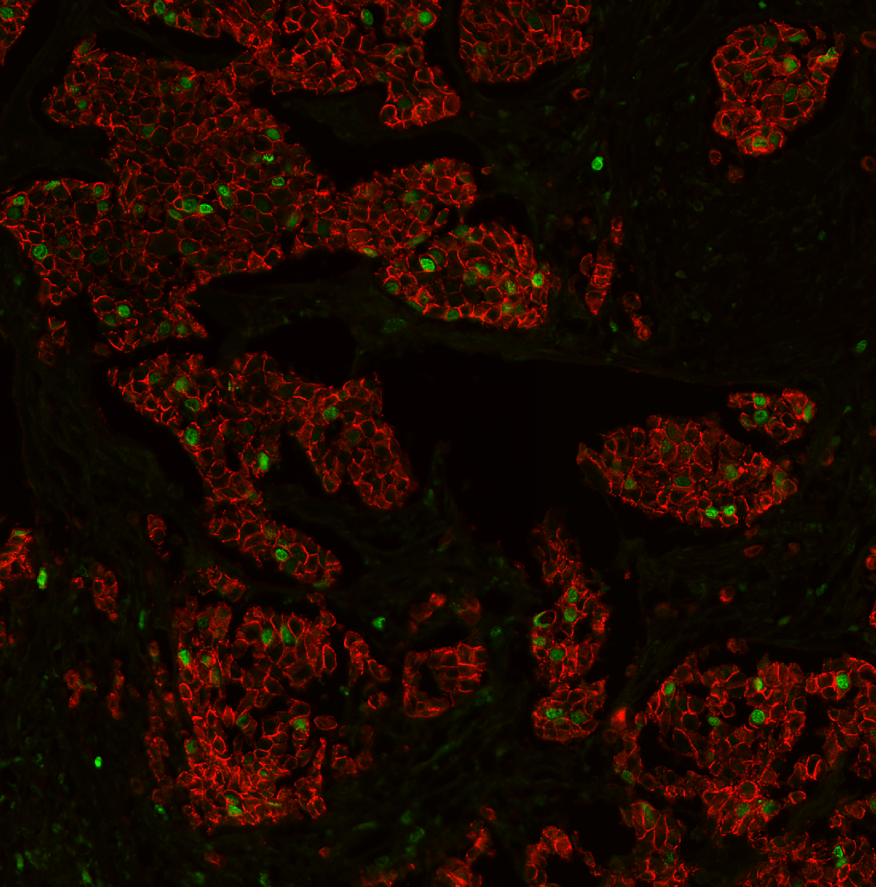
Sarpeda Plex — Marker Separation
Breast tissue: E-cadherin and Ki-67 labeled with the same fluorophore, then separated by AI. Green nuclei (Ki-67) clearly emerge from the red membrane staining (E-cadherin).
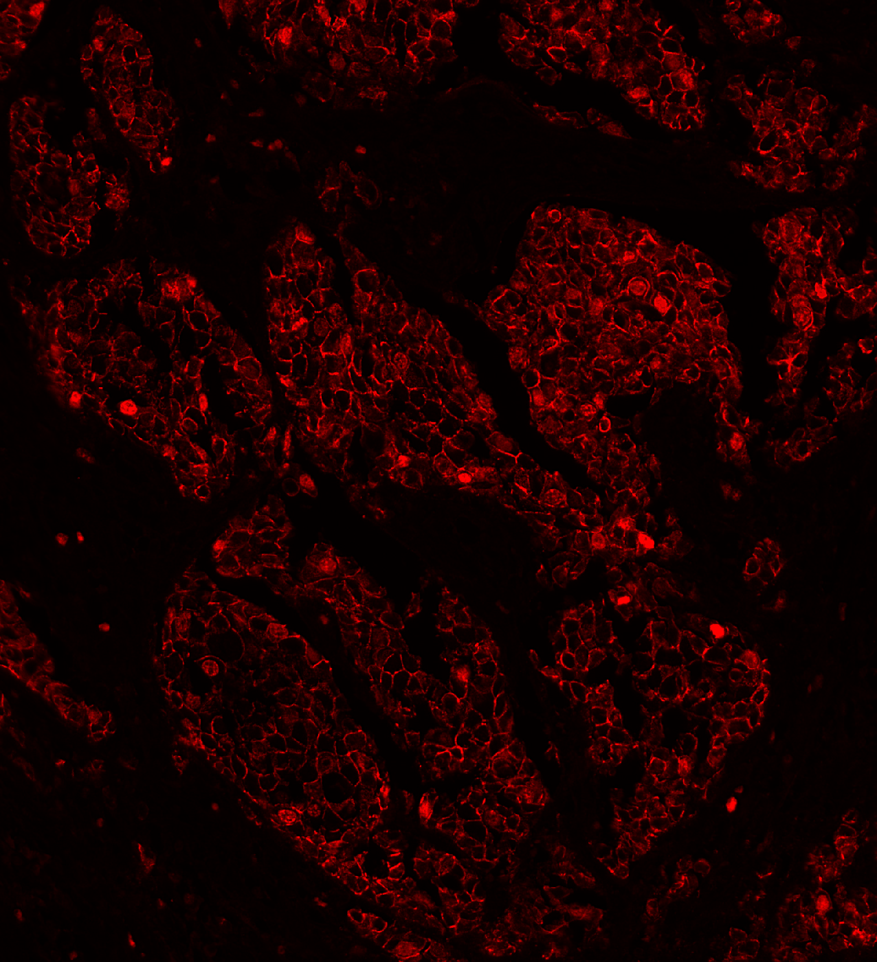
Training image — markers imaged in separate conventional channels
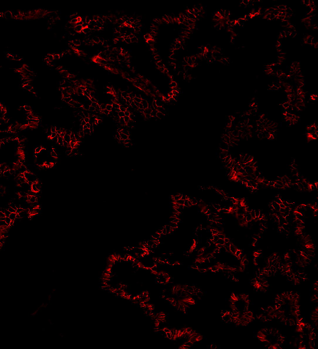
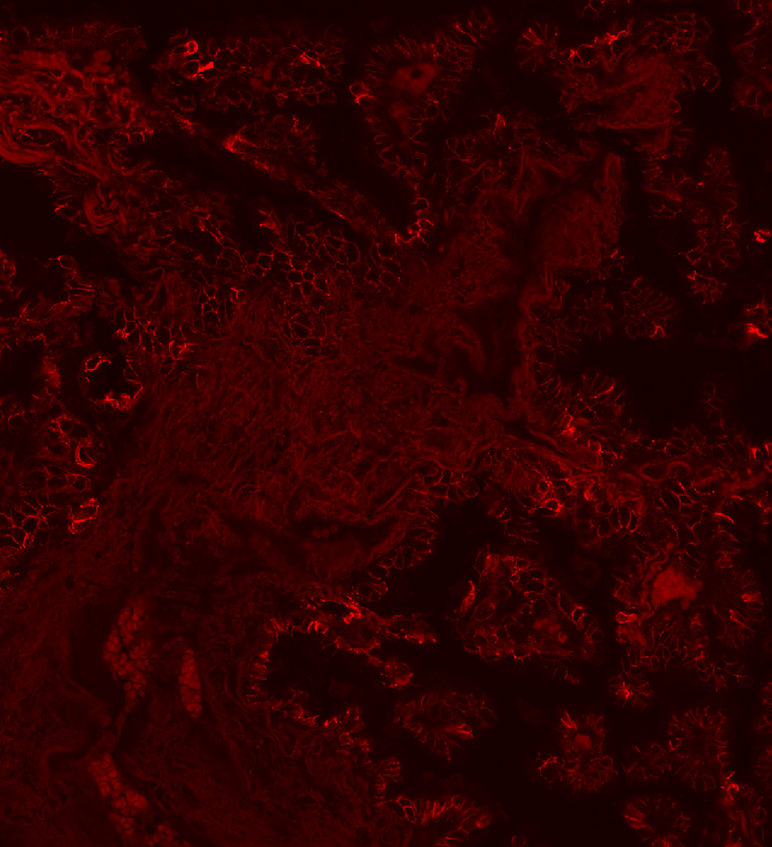
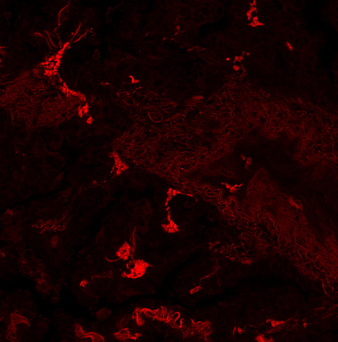
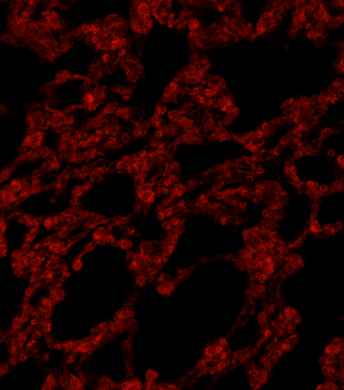
Sarpeda Clear — Background Removal
Lung tissue: E-cadherin signal buried in autofluorescence, then cleaned by AI. The true biological signal emerges with single-pixel detail preserved.
Negative control — background-only reference used for AI training
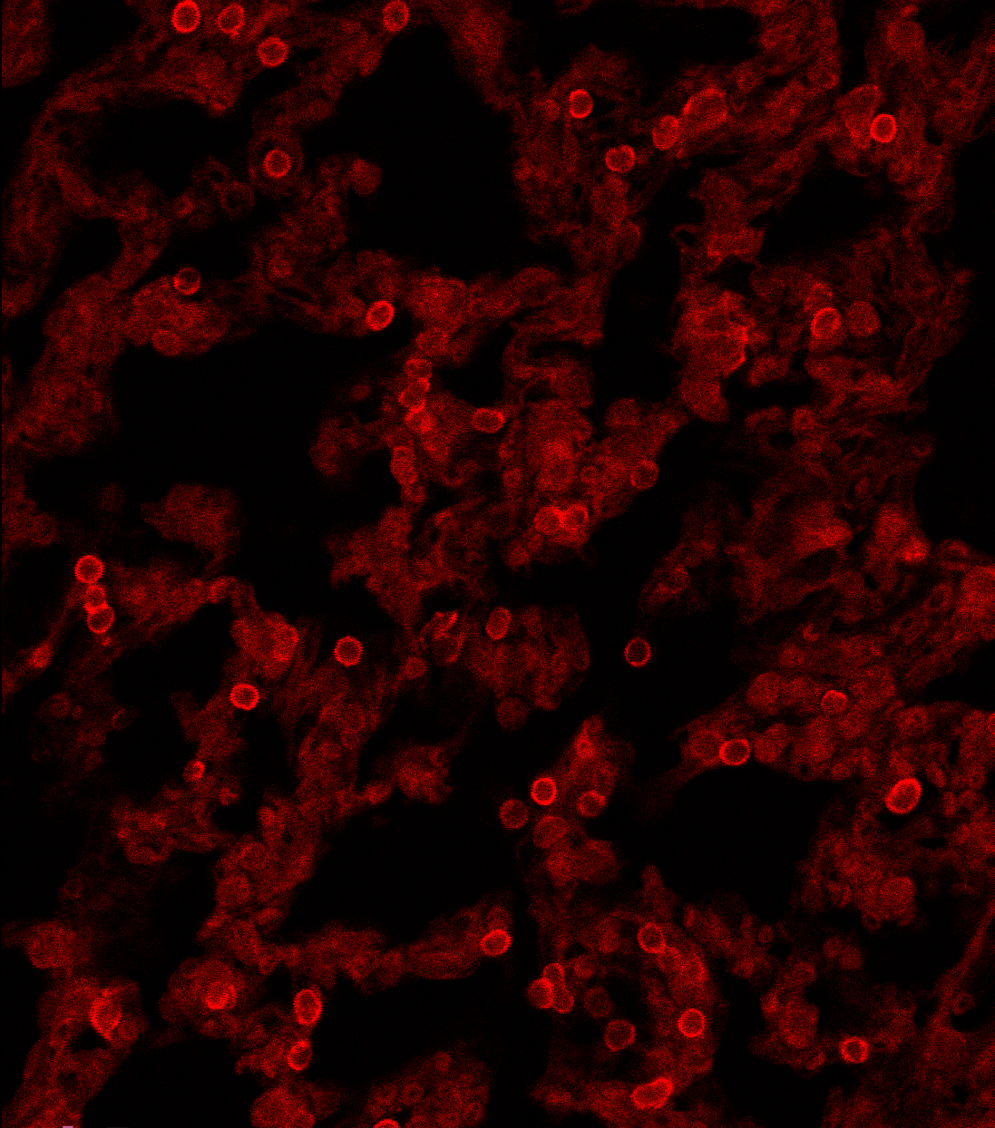
Sarpeda Clear — Crosstalk Removal
Lung tissue, 568 nm channel: the target aSMA signal is contaminated by crosstalk bleeding in from the Rb in the adjacent 647 nm channel. Sarpeda Clear removes the bleed-through and recovers the true aSMA signal.
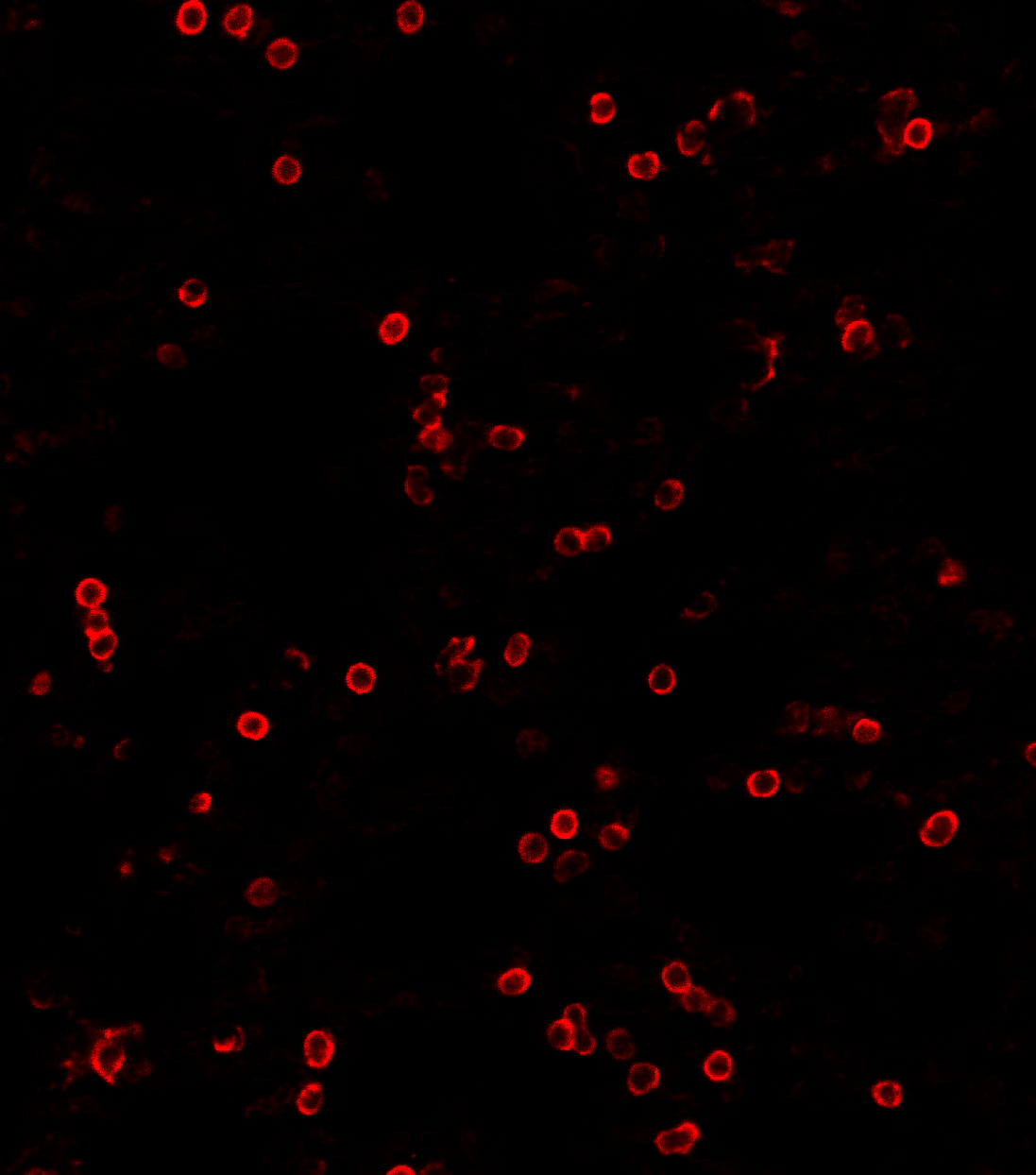
Negative control — background-only reference used for AI training